Call us to ask about same-day availability!
Typical Cases
Oral diseases are a very common finding in pet's mouths. In fact, periodontal disease is the most common disease in cats and dogs over the age of three. Unfortunately, oral diseases are also one of the most under-diagnosed problems in veterinary medicine. These issues are very destructive to the oral soft tissue, bone, and are very painful. Don't expect your pet to show you signs that they have oral pain. It is well known that pets are very good at masking signs of pain. When they are faced with a decision to continue to eat or starve, they almost always choose to keep eating. Below are some pictures of a few cases I've treated. A description will follow the pictures.
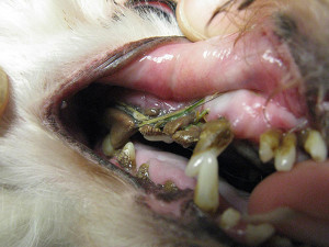
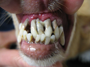
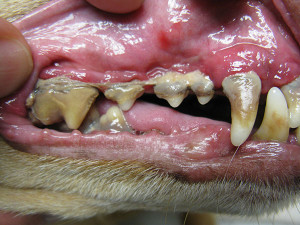
These 3 pictures are of severe periodontal disease. All teeth pictured required extractions due to severe tissue and bone destruction. At their 2 week, post surgical recheck, all patients were doing great. Their owners reported that they were very happy, eating great, and acting like younger dogs. A lot of people believe their pets are less active because they are aging. Living with chronic disease and infection can cause depression, lethargy and inactivity.
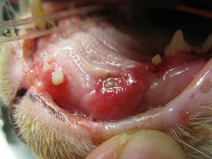
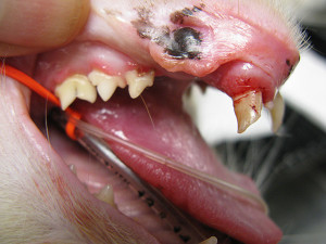
These are pictures of a cat with multiple issues. The picture on the left shows severe gingivitis, a fractured canine tooth below the gingiva that is abscessed. The picture on the right shows a fractured mandibular canine, severe gingivitis, and teeth with root exposure. This cat had full mouth extractions due to end stage periodontal disease. Since his surgery, he only eats dry food and has gained weight. The owners report a much happier and more affectionate cat.
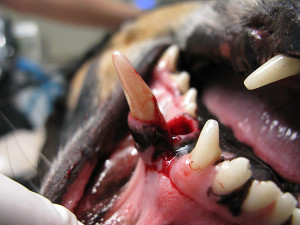
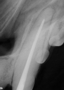
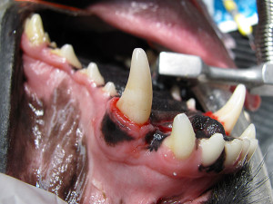
On the left is a picture of a luxated canine tooth from a dog fight. The tooth was saved, but needed a few different procedures. The first step was to replace the tooth into its normal position. The lacerated gingiva was sutured and a fixed implant was placed in the dog's mouth to keep the tooth from moving and to allow the fractured maxilla to heal. Thirty days later, the implant was removed and a root canal was performed. The x-ray shows what the inside of the tooth looks like after a root canal. The picture on the right is of the tooth following the root canal. The dog did great and his 6 month follow up x-rays showed that the root canal was a success.
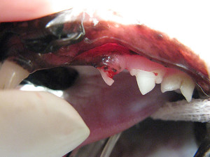
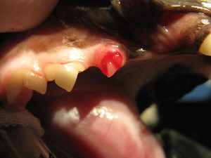
These are two pictures of cats with a very painful process called tooth resorption. Tooth resorption is a destructive process that usually begins in the root(s) of teeth and progresses into the crown. Once the process reaches the crown, it destroys the hard surface of the tooth exposing the nerve endings and blood vessels. No one knows what causes tooth resorption and we cannot halt the progression once it starts. The only treatment available is to surgically remove the painful tooth. Tooth resorption occurs in dogs and cats, but is much more common in cats. Studies have shown that tooth resorption is present in 60% of all cats! That means 6 out of every 10 cats right now have at least one tooth affected!
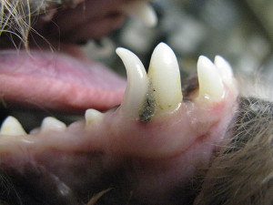
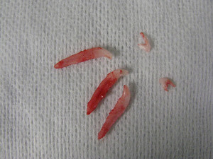
This is a very common problem seen in young dogs, particularly small breeds. This is called retained deciduous teeth or failure to lose their baby teeth. When this occurs, the baby teeth must be extracted. They are occupying a space that is only made for one tooth and cause crowding and malocclusion. If they are not extracted, they will jeopardize the health of the permanent tooth. The picture on the right shows how big the roots of these teeth are. Not only are they large, but they are very fragile. It is common for the root to break during the extraction process. Sometimes it requires oral surgery to remove the complete tooth. Leaving broken roots behind is not an option.
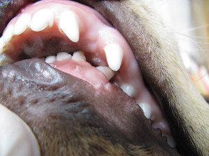
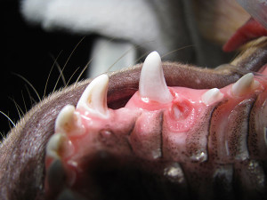
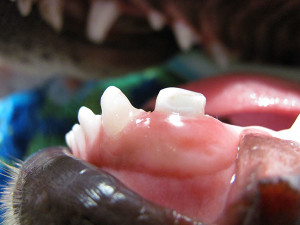
This is a condition called mandibular distocclusion - the mandible is shorter than the maxilla. If the condition is severe enough to cause palatal trauma, it needs to be corrected. Options to correct the trauma and associated pain include: orthodontics, crown reduction with vital pulpotomy (restoration), and extracting the teeth causing pain/trauma. These pictures show the significant trauma caused by the malocclusion and the correction with crown reduction and vital pulpotomy.
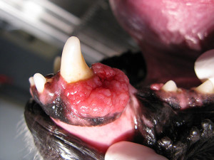
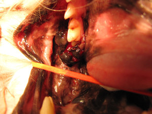
ORAL TUMORS/CANCER
The picture on the left is of a dog with a tumor called acanthomatous ameloblastoma around a mandibular canine tooth. This type of cancer does not metastasize, but is very aggressive locally. Surgery with clean margins is curative. A partial mandibulectomy was performed and the dog shows no signs of recurrence 18 months later. The second picture is of a much more aggressive type of oral cancer called oral melanoma. This dog had major oral surgery (entire mandible on one side removed) followed by 5 weekly Melanoma vaccines. She has had no issues eating and the owners are thrilled with the outcome. She is already months past her life expectancy had she not had surgery.
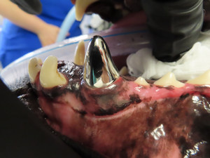
This is a titanium crown that was placed on a dog's canine tooth. Crowns are used to protect damaged teeth and to strengthen teeth after getting a root canal.
Cookies on this website are used to both support the function and performance of the site, and also for marketing purposes, including personalizing content and tailoring advertising to your interests. To manage marketing cookies on this website, please select the button that indicates your preferences. More information can be found in our privacy policy here.

